
phospho-PAK1(S144)+PAK2(S141)+PAK3(S154) Rabbit mAb[9Z27]Cat NO.: A44283
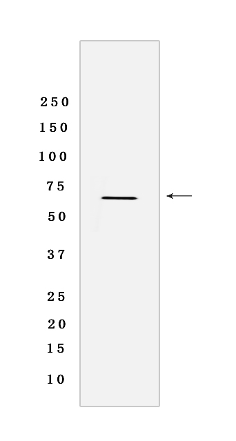
Western blot analysis of extracts from MCF-7 cells lysates EGF-treated .using PAK1(S144)+PAK2(S141)+PAK3(S154) Rabbit mAb[9Z27] at dilution of 1:1000 incubated at 4℃ over night
Product information
Protein names :PAK1,PAK1_HUMAN,Serine/threonine-protein kinase PAK 1
UniProtID :Q13153
MASS(da) :60,647
MW(kDa) :66 kDa
Form :Liquid
Purification :Protein A purification
Host :Rabbit
Isotype :IgG
sensitivity :Endogenous
Reactivity :Human,Mouse,Rat
- ApplicationDilution
- 免疫印迹(WB)1:1000-2000
- 免疫组化(IHC)1:100,
- 免疫荧光(ICC/IF)1:100,
- The optimal dilutions should be determined by the end user
Specificity :Antibody is produced by immunizing animals with a synthetic phosphopeptide corresponding to residues around S144 of Human PAK1.
Storage :Antibody store in 10 mM PBS, 0.5mg/ml BSA, 50% glycerol. Shipped at 4°C. Store at-20°C or -80°C. Products are valid for one natural year of receipt.Avoid repeated freeze / thaw cycles.
WB Positive detected :MCF-7 cells lysates EGF-treated
Function : Protein kinase involved in intracellular signaling pathways downstream of integrins and receptor-type kinases that plays an important role in cytoskeleton dynamics, in cell adhesion, migration, proliferation, apoptosis, mitosis, and in vesicle-mediated transport processes (PubMed:11896197, PubMed:30290153). Can directly phosphorylate BAD and protects cells against apoptosis. Activated by interaction with CDC42 and RAC1. Functions as GTPase effector that links the Rho-related GTPases CDC42 and RAC1 to the JNK MAP kinase pathway. Phosphorylates and activates MAP2K1, and thereby mediates activation of downstream MAP kinases. Involved in the reorganization of the actin cytoskeleton, actin stress fibers and of focal adhesion complexes. Phosphorylates the tubulin chaperone TBCB and thereby plays a role in the regulation of microtubule biogenesis and organization of the tubulin cytoskeleton. Plays a role in the regulation of insulin secretion in response to elevated glucose levels. Part of a ternary complex that contains PAK1, DVL1 and MUSK that is important for MUSK-dependent regulation of AChR clustering during the formation of the neuromuscular junction (NMJ). Activity is inhibited in cells undergoing apoptosis, potentially due to binding of CDC2L1 and CDC2L2. Phosphorylates MYL9/MLC2. Phosphorylates RAF1 at
Tissue specificity :Overexpressed in gastric cancer cells and tissues (at protein level) (PubMed:25766321)..
Subcellular locationi :Cytoplasm. Cell junction, focal adhesion. Cell projection, lamellipodium. Cell membrane. Cell projection, ruffle membrane. Cell projection, invadopodium. Nucleus, nucleoplasm. Chromosome. Cytoplasm, cytoskeleton, microtubule organizing center, centrosome.
IMPORTANT: For western blots, incubate membrane with diluted primary antibody in 1% w/v BSA, 1X TBST at 4°C overnight.
-
About
-
Product
-
Law
-
Literature
-

微信扫码关注我们